POSTPARTUM COMPLICATIONS (MATERNAL REVIEWER)
1. POSTPARTAL HEMORRHAGE
Hemorrhage, one of the most important causes of maternal mortality associated with childbearing, poses a possible threat throughout pregnancy and is also a major potential danger in the immediate postpartum period.
• Postpartum hemorrhage has been defined as any blood loss from the uterus greater than 500 mL within 24 hours.
• In specific agencies, the loss may not be considered hemorrhage until it reaches 1000 mL. Hemorrhage may occur either early (within the first 24 hours) or late (any time after the first 24 hours during the remaining days of the 6-week puerperium).
• The greatest danger of hemorrhage is in the first 24 hours because of the grossly denuded and unprotected uterine area left after detachment of the placenta.
• There are five main causes for postpartum hemorrhage: uterine atony, lacerations, retained placental fragments, uterine inversion, and disseminated intravascular coagulation.
A. Uterine Atony
Uterine atony, or relaxation of the uterus, is the most frequent cause of postpartum hemorrhage. The uterus must remain in a contracted state after birth to keep the open vessels at the placental site from bleeding. If the uterus suddenly relaxes, there will be an abrupt gush of blood vaginally from the placental site. If the vaginal bleeding is extremely copious, a woman will exhibit symptoms of shock and blood loss. This can occur immediately after birth or more gradually, over the first postpartum hour, as the uterus slowly becomes uncontracted. It is difficult to estimate the amount of blood a postpartum woman has lost, because it is difficult to estimate the amount of blood it takes to saturate a perineal pad. The amount is between 25 and 50 mL. Factors that predispose to poor uterine tone or any inability to maintain a contracted state are:
• Deep anesthesia or analgesia
• Labor initiated or assisted with an oxytocin agent
• Maternal age greater than 35 years
• High parity
• Previous uterine surgery
• Prolonged and difficult labor
• Possible chorioamnionitis Secondary maternal illness (e.g., anemia)
• Prior history of postpartum hemorrhage Endometritis
• Prolonged use of magnesium sulfate or other tocolytic therapy
Therapeutic Management:
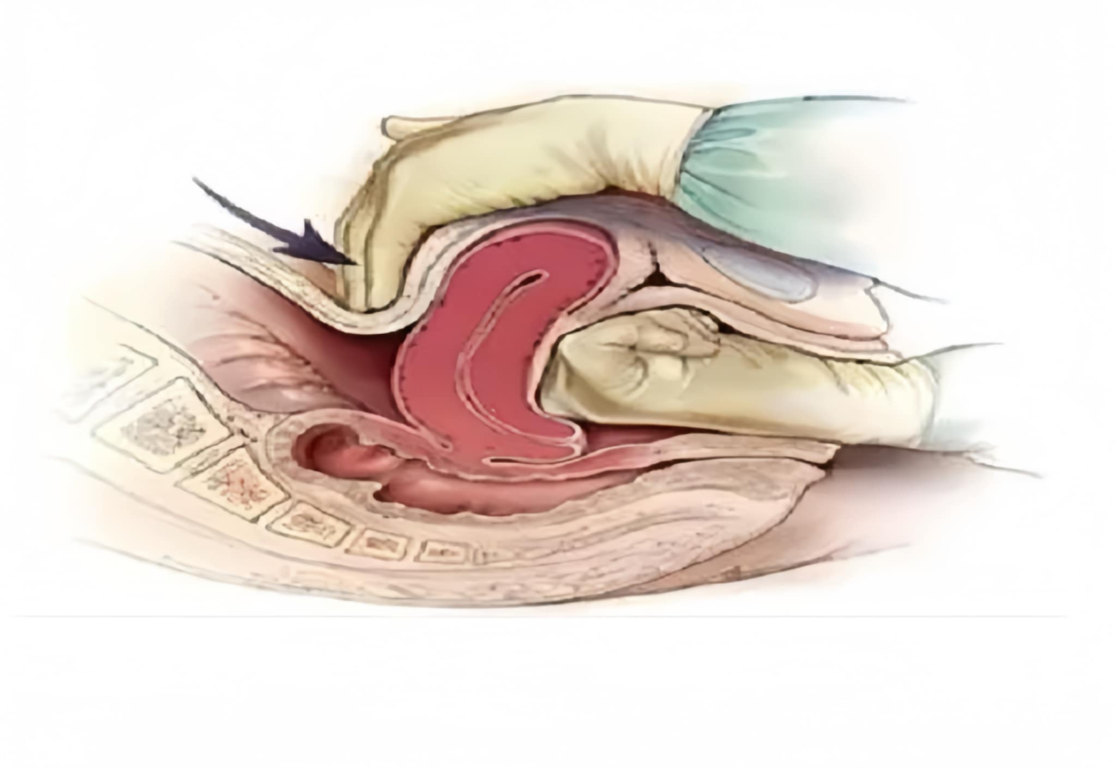
1. Bimanual Massage
If fundal massage and administration of oxytocin or methylergonovine are not effective in stopping uterine bleeding, a sonogram may be done to detect possible retained placental fragments. The woman’s physician or nurse- midwife may attempt bimanual compression. With this procedure, the physician or nurse-midwife inserts one hand into a woman’s vagina while pushing against the fundus through the abdominal wall with the other hand.
2. Prostaglandin Administration
Prostaglandins promote strong, sustained uterine contractions. Intramuscular injection of prostaglandin F22 is another way to initiate uterine contractions. Carboprost tromethamine (Hemabate), a prostaglandin F2a derivative, or methylergonovine maleate (Methergine), an ergot compound, given intramuscularly, are second possibilities. Rectal misoprostol, a prostaglandin E1 analogue, may be administered rectally. Hemabate may be repeated every 15 to 90 minutes up to 8 doses; methylergonovine may be repeated every 2 to 4 hours up to 5 doses. The usual dosage of oxytocin is 10 to 40 U per 1000 mL of a Ringer’s lactate solution. When oxytocin is given intravenously, its action is immediate 3.
3. Blood Replacement
Blood transfusion to replace blood loss with postpartum hemorrhage may be necessary. Make certain that blood typing and cross-matching were done when the woman was admitted, and that blood is available. Women who experience postpartum hemorrhage tend to have a longer than average recovery period, because the physiologic exhaustion of body systems can interfere with their recovery. Iron therapy may be prescribed to ensure good hemoglobin formation. Activity level, exertion, and postpartum exercise may be restricted somewhat.
• Monitor her temperature closely in the postpartum period, to detect the earliest signs of developing infection.
4. Hysterectomy or Suturing
Usually, therapeutic management is effective in halting bleeding. In the rare instance of extreme uterine atony, sutures or balloon compression may be used to halt bleeding. Embolization of pelvic and uterine vessels by angiographic techniques may be successful. As a last resort, ligation of the uterine arteries or a hysterectomy may be necessary. Open lines of communication between the couple and health care providers that allow a family to vent its feelings are most helpful to a couple in this crisis.
B. Lacerations
Small lacerations or tears of the birth canal are common and may be considered a normal consequence of childbearing. Large lacerations, however, can cause complications.
They occur most often:
• With difficult or precipitate births
• In primigravida
• With the birth of a large infant ( 9 lb)
• With the use of a lithotomy position and instruments
1. Cervical Lacerations
Lacerations of the cervix are usually found on the sides of the cervix, near the branches of the uterine artery. If the artery is torn, the blood loss may be so great that blood gushes from the vaginal opening. Because this is arterial bleeding, it is brighter red than the venous blood lost with uterine atony. Repair of a cervical laceration is difficult because the bleeding can be so intense that it obstructs visualization of the area. Be certain that a physician or nurse-midwife has adequate space to work, adequate sponges and suture supplies, and a good light source. If the cervical laceration appears to be extensive or difficult to repair, it may be necessary for the woman to be given a regional anesthetic to relax the uterine muscle and to prevent pain.
2. Vaginal Lacerations
Although they are rare, lacerations can also occur in the vagina. They are easier to assess than cervical lacerations because they are easier to view. Because vaginal tissue is friable, vaginal lacerations are also hard to repair. Some oozing often occurs after a repair, so the vagina may be packed to maintain pressure on the suture line. If packing is inserted, document in a woman’s nursing care plan when and where it was placed, so you can be certain it will be removed after 24 to 48 hours or before discharge. An indwelling urinary catheter (Foley catheter) may be placed at the same time because the packing causes pressure on the urethra and can interfere with voiding.
3. Perineal Lacerations
Lacerations of the perineum usually occur when a woman is placed in a lithotomy position for birth, because this position increases tension on the perineum. Perineal lacerations are sutured and treated as an episiotomy repair. Make certain that the degree of the laceration is documented, because women with fourthdegree lacerations need extra precautions to avoid having repair sutures loosened or infected. C. Retained Placental Fragments
Occasionally, a placenta does not deliver in its entirety; fragments of it separate and are left behind. Because the portion retained keeps the uterus from contracting fully, uterine bleeding occurs. To detect the complication of retained placenta, every placenta should be inspected carefully after birth to see that it is complete. Retained placental fragments may also be detected by ultrasound. A blood serum sample that contains human chorionic gonadotropin hormone (hCG) also reveals that part of a placenta is still present. Removal of the retained placental fragment is necessary to stop the bleeding.
D. Disseminated Intravascular Coagulation
Disseminated intravascular coagulation (DIC) is a deficiency in clotting ability caused by vascular injury. It may occur in any woman in the postpartum period, but it is usually associated with premature separation of the placenta, a missed early miscarriage, or fetal death in utero.
E. Subinvolution
Subinvolution is incomplete return of the uterus to its prepregnant size and shape. With subinvolution, at a 4- or 6week postpartum visit, the uterus is still enlarged and soft. Lochia discharge usually is still present. Subinvolution may result from a small retained placental fragment, a mild endometritis (infection of the endometrium), or an accompanying problem such as a uterine myoma that is interfering with complete contraction.
F. Perineal Hematomas
A perineal hematoma is a collection of blood in the subcutaneous layer of tissue of the perineum. The overlying skin, as a rule, is intact with no noticeable trauma. Such blood collections can be caused by injury to blood vessels in the perineum during birth. They are most likely to occur after rapid, spontaneous births and in women who have perineal varicosities.
2. Puerperal Infection
Infection of the reproductive tract is another leading cause of maternal mortality. When caring for a woman who has any of these circumstances, be aware that the risk for postpartum infection is greatly increased. Theoretically, the uterus is sterile during pregnancy and until the membranes rupture. After rupture, pathogens can invade. The risk of infection is even greater if tissue edema and trauma are present. A puerperal infection is always potentially serious, because, although it usually begins as only a local infection, it can spread to involve the peritoneum (peritonitis) or the circulatory system (septicemia). These conditions can be fatal in a woman whose body is already stressed from childbirth. A. Endometritis

B. Infection of the Perineum
If a woman has a suture line on her perineum from an episiotomy or a laceration repair, a portal of entry exists for bacterial invasion. Infections of the perineum usually remain localized. They are revealed by symptoms like those of any suture-line infection, such as pain, heat, and a feeling of pressure. The woman may or may not have an elevated temperature, depending on the systemic effect and spread of the infection. Inspection of the suture line reveals the inflammation. One or two stitches may be sloughed away, or an area of the suture line may be open with purulent drainage present. Notify the woman’s physician or nurse-midwife of the localized symptoms, and culture the discharge using a sterile cotton-tipped applicator touched to the secretions. A woman’s physician or nurse-midwife may choose to remove perineal sutures, to open the area and allow for drainage. Packing, such as iodoform gauze, may be placed in the open lesion to keep it open and allow drainage. Be sure the woman is aware that the packing is in place, so she knows not to dislodge it as she changes her perineal pad.
C. Peritonitis
Peritonitis, or infection of the peritoneal cavity, usually occurs as an extension of endometritis. It is one of the gravest complications of childbearing and is a major cause of death from puerperal infection. The infection spreads through the lymphatic system or directly through the fallopian tubes or uterine wall to the peritoneal cavity. An abscess may form in the cul-de-sac of Douglas because this is the lowest point of the peritoneal cavity and gravity causes infected material to localize there. Symptoms are the same as those of a surgical patient in whom a peritoneal infection develops rigid abdomen, abdominal pain, high fever, rapid pulse, vomiting, and the appearance of being acutely ill. When assessing the abdomen of a postpartum woman, be sure to note not only that her uterus is well contracted but also that the remainder of her abdomen s soft. The occurrence of a rigid abdomen (guarding) is one of the first symptoms of peritonitis.
D. Thrombophlebitis
Phlebitis is inflammation of the lining of a blood vessel. Thrombophlebitis is inflammation with the formation of blood clots. When thrombophlebitis occurs in the postpartal period, it is usually an extension of an endometrial infection. It tends to occur because:
• A woman’s fibrinogen level is still elevated from pregnancy, leading to
increased blood clotting.
• Dilatation of lower extremity veins is still present as a result of pressure of the
fetal head during pregnancy and birth.
• The relative inactivity of the period or a prolonged time spent in delivery or
birthing room stirrups leads to pooling, stasis, and clotting of blood in the lower
extremities.
• Obesity from increased weight before pregnancy and pregnancy weight gain
can lead to relative inactivity and lack of exercise.
• The woman smokes cigarettes.
Thrombophlebitis is classified as superficial vein disease (SVD) or deep vein thrombosis (DVT). Women most prone to thrombophlebitis are those who are obese, have varicose veins, have had a previous thrombophlebitis, are older than 35 years of age with increased parity, or have a high incidence of thrombophlebitis in their family.
E. Urinary Tract Infection
A woman who is catheterized at the time of childbirth or during the postpartum period is prone to development of a urinary tract infection, because bacteria may be introduced into the bladder at the time of catheterization. If a urinary tract infection develops, a woman notices symptoms of burning on urination, possibly blood in the urine (hematuria), and a feeling of frequency or that she always must void. The pain is so sharp on voiding that she may resist voiding, further compounding the problem of urinary stasis. She may also have a low-grade fever and discomfort from lower abdominal pain.
Obtain a clean-catch urine specimen from any woman with symptoms of urinary tract infection. This can be done as an independent nursing action. So that lochia discharge does not contaminate the specimen, provide a sterile cotton ball for the woman to tuck into her vagina after perineal cleansing. Be certain to ask if she removed the cotton ball after the procedure; otherwise, it could cause stasis of vaginal secretions and increase the possibility of end
Although sulfa drugs are normally prescribed for urinary tract infection, they are contraindicated for breastfeeding women because they can cause neonatal jaundice. Typically, therefore, a broad-spectrum antibiotic such as amoxicillin or ampicillin will be prescribed to treat a postpartum urinary tract infection. If an antibiotic contraindicated by breastfeeding is prescribed, check with a woman’s physician about possibly changing the antibiotic to one that is safe for breastfeeding. Encourage a woman to drink large amounts of fluid (a glass every hour) to help flush the infection from her bladder. She may need an oral analgesic, such as acetaminophen (Tylenol), to reduce the pain of urination for the next few times she voids until the antibiotic begins to have an effect and the burning sensation disappears. Although symptoms of urinary tract infection decrease quickly, be certain a woman understands the importance of continuing to take the prescribed antibiotic for the full 5 to 7 days to eradicate the infection completely.
3. Emotional and Psychological Complications of Puerperium
A. Postpartum Depression
Almost every woman notices some immediate (1 to 10 days postpartum) feelings of sadness (postpartum “blues”) after childbirth. This probably occurs as a response to the anticlimactic feeling after birth and probably is related to hormonal shifts as the levels of estrogen, progesterone, and gonadotropin-releasing hormone in her body decline or rise. The sensations of overwhelming sadness can interfere with breastfeeding, childcare, and returning to work. In addition to an overall feeling of sadness, a woman may notice extreme fatigue, an inability to stop crying, increased anxiety about her own or her infant’s health, insecurity (unwillingness to be left alone or inability to make decisions), psychosomatic symptoms (nausea and vomiting, diarrhea), and either depressive or manic mood fluctuations. Depression of this kind is termed postpartum depression and reflects a more serious problem than normal “baby blues”. Risk factors for postpartum depression include a history of depression, a troubled childhood, low self-esteem, stress in the home or at work, and lack of effective support people. Different expectations between partners or disappointment in the child could play
major roles. It is difficult to predict which women will develop postpartum depression before
birth, because childbirth can result in so many varied reactions; if factors could be identified, pregnancy counseling might be able to prevent symptoms. In the postpartum period, discovery of the problem as soon as symptoms develop nursing priority. Several depression scales to help detect postpartum depression are available but conscientious observation and discussion with women can reveal symptoms just as well. A woman may need counseling and possibly antidepressant therapy to integrate the experience of childbirth into her life. This is crucial to development of a healthy maternal–infant bond, to the health of any other children in the family, and to overall family functioning. Ask at postpartum return visits and well-child visits about symptoms that would suggest depression and recommend an appropriate referral.
B. Postpartum Psychosis
As many as 1 woman in 500 has enough symptoms during the year after the birth of a child to be considered psychiatrically ill. When the illness coincides with the postpartum period, it is called postpartum psychosis. Rather than being a response to the physical aspects of childbearing, however, it is probably a response to the crisis of childbearing. Most of these women have had symptoms of mental illness before pregnancy. If the pregnancy had not precipitated the illness, a death in the family, loss of a job or income, divorce, or some other major life crisis would probably have precipitated the same recurrence. A woman with postpartum psychosis usually appears exceptionally sad. Psychosis exists when a person has lost contact with reality. A woman with a postpartum psychosis may deny that she has had a child and, when the child is brought to her, insist that she was never pregnant. She may voice thoughts of infanticide or that her infant is possessed. If observation tells you that a woman is not functioning, you cannot improve her concept of reality by a simple measure such as explaining what a correct perception is. Her sensory input is too disturbed to comprehend this. In addition, she may interpret your attempt as threatening. She may respond with anger or become equally threatening. A psychosis is a severe mental illness that requires referral to a professional psychiatric counselor and antipsychotic medication. Do not leave the woman alone, because her distorted perception might lead her to harm herself. Nor should you leave her alone with her infant.
 POSTPARTUM COMPLICATIONS (MATERNAL REVIEWER)


Comments
Post a Comment